Home

ciliegia sono fiero negativo staining intensity principio Condividere Adatto

FR-β staining intensity correlations. IHC was performed on a BioMax... |  Download Scientific Diagram
FR-β staining intensity correlations. IHC was performed on a BioMax... | Download Scientific Diagram
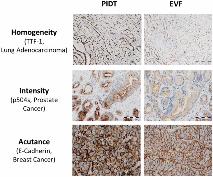
Critical assessment of staining properties of a new visualization  technology: a novel, rapid and powerful immunohistochemical detection  approach | Histochemistry and Cell Biology
Critical assessment of staining properties of a new visualization technology: a novel, rapid and powerful immunohistochemical detection approach | Histochemistry and Cell Biology

Mesure color intensity of DAB image - Image Analysis - Image.sc Forum

Biomedicines | Free Full-Text | Immunohistochemical Evaluation of Candidate  Biomarkers for Fluorescence-Guided Surgery of Myxofibrosarcoma Using an  Objective Scoring Method
Biomedicines | Free Full-Text | Immunohistochemical Evaluation of Candidate Biomarkers for Fluorescence-Guided Surgery of Myxofibrosarcoma Using an Objective Scoring Method

Figures and data in Highly multiplexed immunofluorescence imaging of human  tissues and tumors using t-CyCIF and conventional optical microscopes |  eLife
Figures and data in Highly multiplexed immunofluorescence imaging of human tissues and tumors using t-CyCIF and conventional optical microscopes | eLife
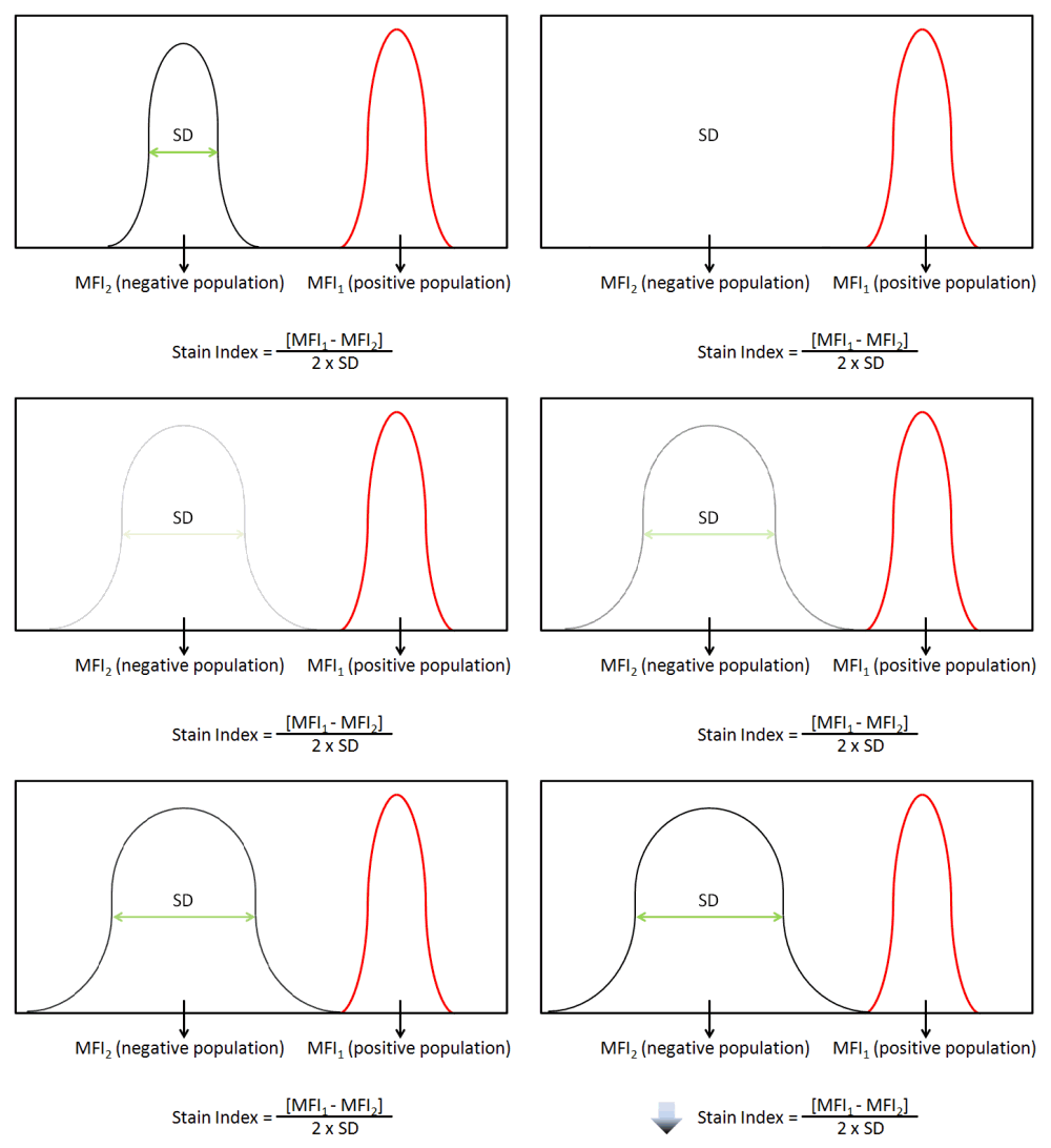
Blog - The Stain Index: What Is It and What Does It Tell You?

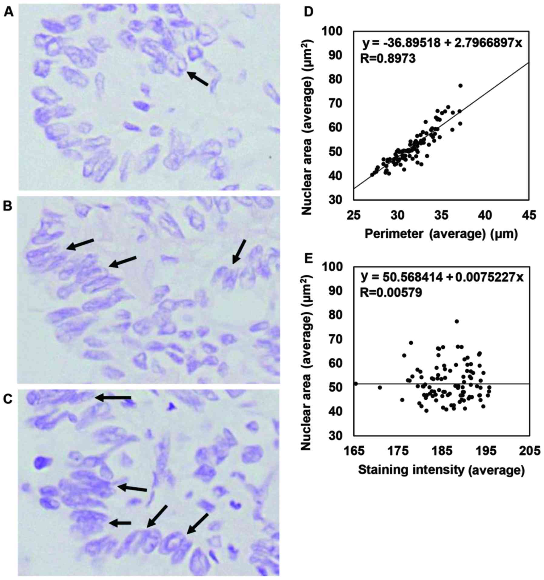
Image analysis of the nuclear characteristics of emerin protein and the  correlation with nuclear grooves and intranuclear cytoplasmic inclusions in  lung adenocarcinoma
Image analysis of the nuclear characteristics of emerin protein and the correlation with nuclear grooves and intranuclear cytoplasmic inclusions in lung adenocarcinoma
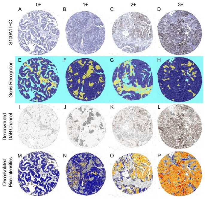
Quantitative comparison of immunohistochemical staining measured by digital  image analysis versus pathologist visual scoring | Diagnostic Pathology |  Full Text
Quantitative comparison of immunohistochemical staining measured by digital image analysis versus pathologist visual scoring | Diagnostic Pathology | Full Text

How to measure the staining intensity without positive cell detection -  Image Analysis - Image.sc Forum
How to measure the staining intensity without positive cell detection - Image Analysis - Image.sc Forum

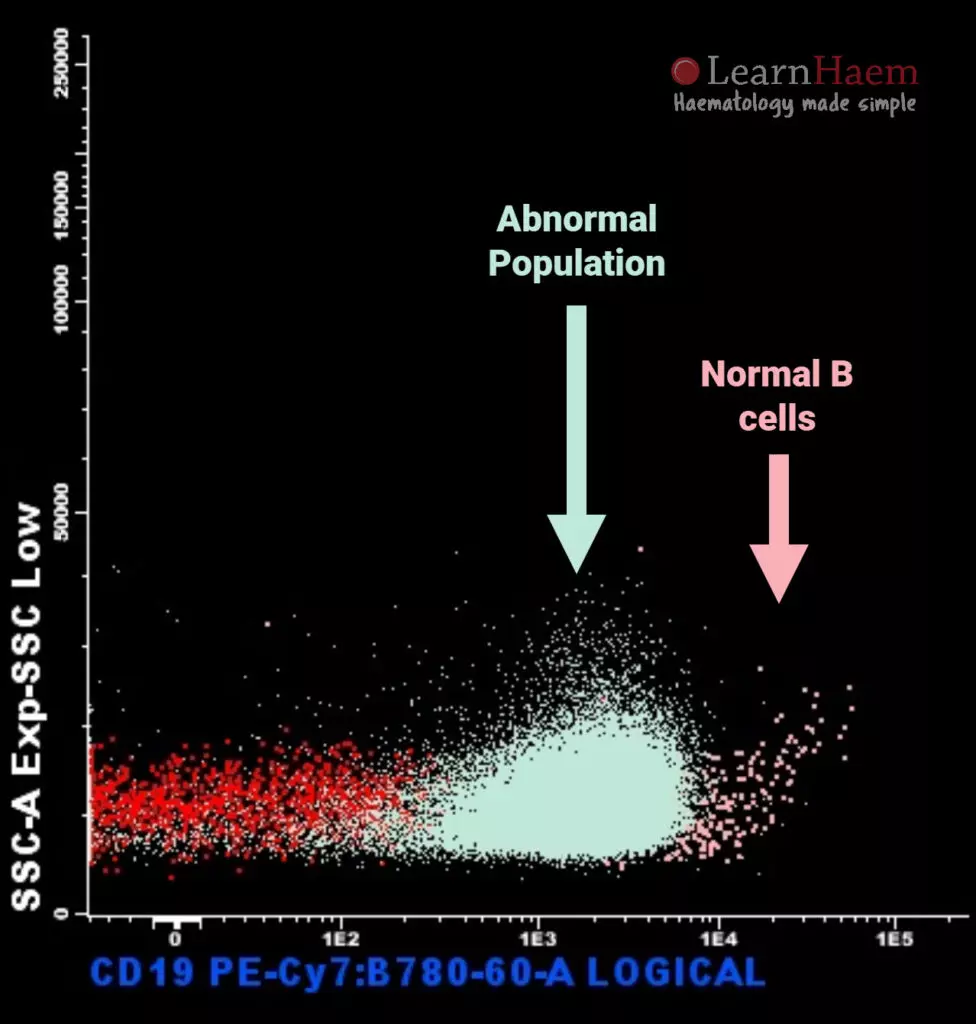
Reporting - LearnHaem | Haematology Made Simple

Critical assessment of staining properties of a new visualization  technology: a novel, rapid and powerful immunohistochemical detection  approach | Histochemistry and Cell Biology
Critical assessment of staining properties of a new visualization technology: a novel, rapid and powerful immunohistochemical detection approach | Histochemistry and Cell Biology
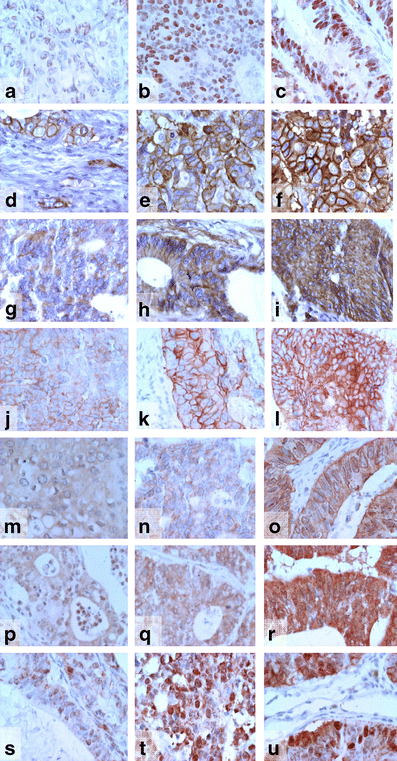
Value of staining intensity in the interpretation of immunohistochemistry  for tumor markers in colorectal cancer | Virchows Archiv
Value of staining intensity in the interpretation of immunohistochemistry for tumor markers in colorectal cancer | Virchows Archiv

FalseColor-Python: A rapid intensity-leveling and digital-staining package  for fluorescence-based slide-free digital pathology | PLOS ONE
FalseColor-Python: A rapid intensity-leveling and digital-staining package for fluorescence-based slide-free digital pathology | PLOS ONE
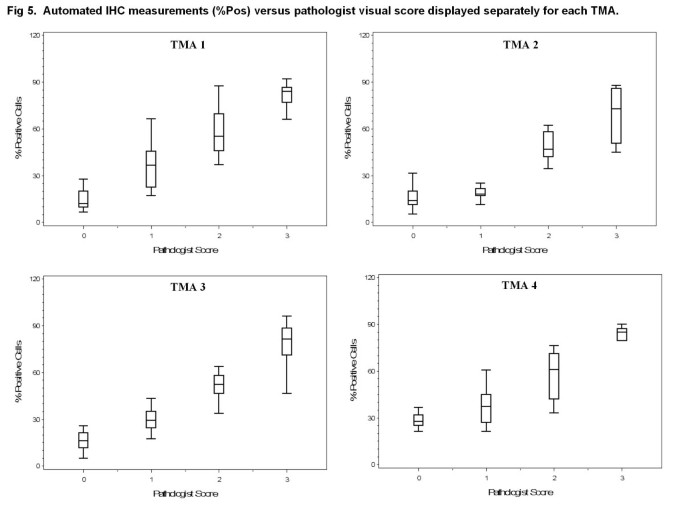
Quantitative comparison of immunohistochemical staining measured by digital  image analysis versus pathologist visual scoring | Diagnostic Pathology |  Full Text
Quantitative comparison of immunohistochemical staining measured by digital image analysis versus pathologist visual scoring | Diagnostic Pathology | Full Text

Using ImageJ to measure the mean staining intensity of two different images

Staining intensity for IHC methods in sections of different thicknesses...  | Download Scientific Diagram
Staining intensity for IHC methods in sections of different thicknesses... | Download Scientific Diagram

HER2 staining intensity in HER2-positive disease: relationship with FISH  amplification and clinical outcome in the HERA trial of adjuvant  trastuzumab - ScienceDirect
HER2 staining intensity in HER2-positive disease: relationship with FISH amplification and clinical outcome in the HERA trial of adjuvant trastuzumab - ScienceDirect

SOX2 IHC staining intensity is high in CRC tissues. (A) The staining... |  Download Scientific Diagram
SOX2 IHC staining intensity is high in CRC tissues. (A) The staining... | Download Scientific Diagram
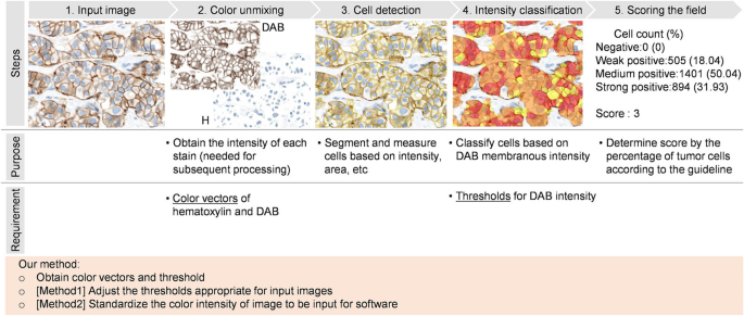
Analysis of immunohistochemistry staining intensity using... | Download  Scientific Diagram
Analysis of immunohistochemistry staining intensity using... | Download Scientific Diagram

How to measure the staining INTENSITY of NUCLEUS and CYTOPLASM using ImageJ  Software
How to measure the staining INTENSITY of NUCLEUS and CYTOPLASM using ImageJ Software

Standardizing HER2 immunohistochemistry assessment: calibration of color  and intensity variation in whole slide imaging caused by staining and  scanning | Applied Microscopy | Full Text
Standardizing HER2 immunohistochemistry assessment: calibration of color and intensity variation in whole slide imaging caused by staining and scanning | Applied Microscopy | Full Text

PELVIPHARM - Protein expression – Immunohistochemistry - Immunofluorescence

PDF] Comparison of Special Stains for Keratin with Routine Hematoxylin and  Eosin Stain | Semantic Scholar
PDF] Comparison of Special Stains for Keratin with Routine Hematoxylin and Eosin Stain | Semantic Scholar

Mesure color intensity of DAB image - Image Analysis - Image.sc Forum